

What is CSF Shunt Procedure
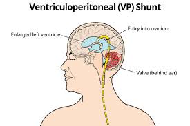
A ventriculoperitoneal (VP) shunt is a gadget used to alleviate weight from the mind brought about by liquid aggregation. VP shunting is a surgical strategy that is fundamentally used to treat a condition called hydrocephalus, which happens when abundance cerebrospinal liquid (CSF) gathers in the cerebrum's ventricles.
What are the types?
CSF pads your mind and shields it from damage inside your skull. The liquid goes about as a conveyance framework for supplements that your cerebrum needs, furthermore takes away waste items. Typically, CSF courses through these ventricles to the base of the cerebrum. The liquid then showers the cerebrum and spinal rope before it is reabsorbed into the blood. At the point when this ordinary stream is upset, the development of liquid can make hurtful weight on the brain's tissues, which can harm the cerebrum. VP shunts are surgically set inside one of the cerebrum's ventricles to redirect liquid far from the mind and restore typical stream and ingestion of CSF.
Sign and Symptoms
Blockages are the most common cause of hydrocephalus. Cysts, tumors, or inflammation in the brain can impede the normal flow of CSF, creating an unsafe accumulation. Symptoms of hydrocephalus can include: • large head size • headaches • seizures • irritability • loss of previously acquired skills • incontinence • poor appetite • cognitive delays or regression • memory loss • poor coordination • impaired vision
Treatment Procedure
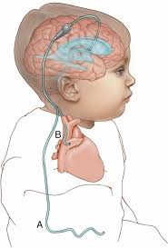
The surgical procedure to implant a VP (ventricular peritoneal) shunt usually requires less than an hour in the operating room. After the patient is placed under general anesthesia. Incisions are then made on the head and in the abdomen to allow the neurosurgeon to pass the shunt's tubing through the fatty tissue just under the skin. A small hole is made in the skull, opening the membranes between the skull and brain to allow the ventricular end of the shunt to be passed through the brain and into the lateral ventricle. The abdominal (peritoneal) end is passed into the abdominal cavity through a small opening in the lining of the abdomen where the excess CSF will eventually be absorbed. The incisions are then closed and sterile bandages are applied.